Knee Surgery
Home » Operations » Knee Surgery
Cruciate Ligament Surgery
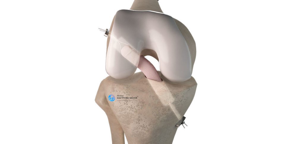
4-Strand Semitendinosus Tendon ACL Reconstruction
Anterior cruciate ligament rupture results in knee instability or the premature wear of the menisci, a potential source of early osteoarthritis in young patients. Arthroscopic reconstruction of the anterior cruciate ligament using a short graft of the semitendinosus tendon (4-strand semitendinosus autograft) is a reliable, proven method for stabilising the knee and enabling a return to the same level of sports activity as before. This technique has the advantage of using only one tendon with excellent mechanical strength and very low donor site morbidity.
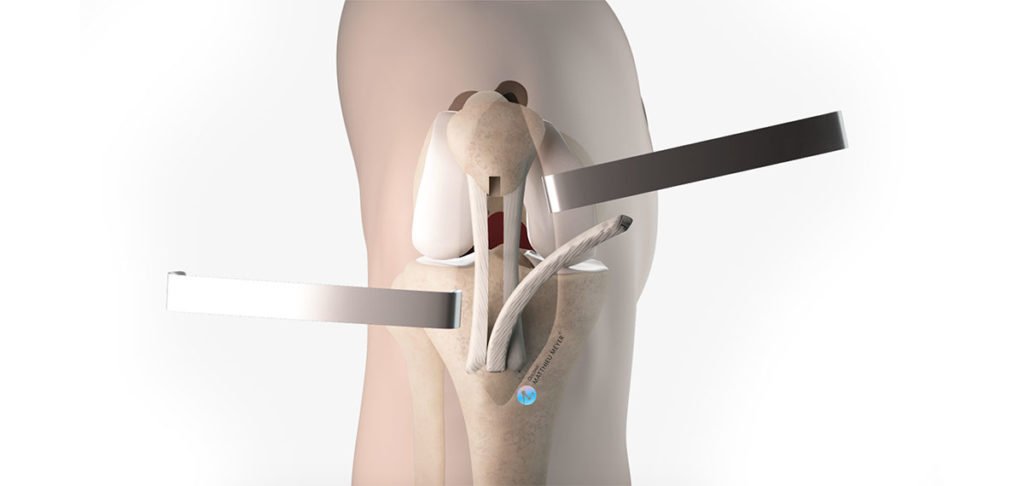
Anterior Cruciate Ligament Reconstruction (Kenneth-Jones)
Anterior cruciate ligament rupture results in knee instability or the premature wear of the menisci, a potential source of early osteoarthritis in young patients. Arthroscopic reconstruction of the anterior cruciate ligament using a bone-tendon-bone graft of the patellar tendon (Kenneth-Jones technique) is a tried and tested method for stabilising the knee and enabling a return to the same level of sports activity as before. This technique is often a cause of transitory anterior discomfort around the harvest site of the tendon graft and is currently used as a second-line treatment when the hamstring tendons have already been harvested.
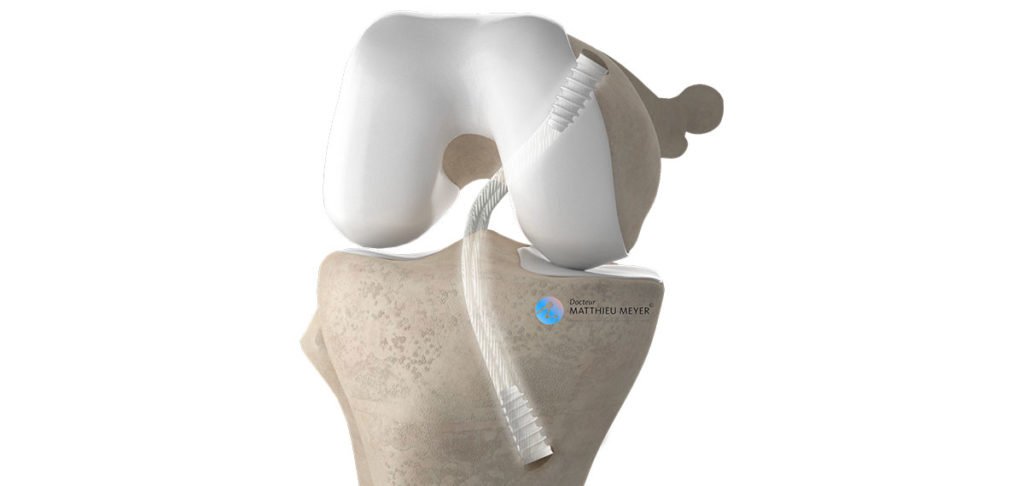
Posterior Cruciate Ligament Reconstruction
Posterior cruciate ligament rupture is rare and is caused by a direct trauma on the anterior side of the knee. If the peripheral ligaments are involved, it is essential posterior cruciate ligament reconstruction is performed relatively urgently. However, with an isolated posterior cruciate ligament tear, reconstruction is only necessary in the chronic phase in the case of knee instability or patellar pain. Arthroscopic posterior cruciate ligament reconstruction is achieved using a tendon autograft or in some cases, rapidly after the rupture, using a synthetic graft (LARS artificial ligament).
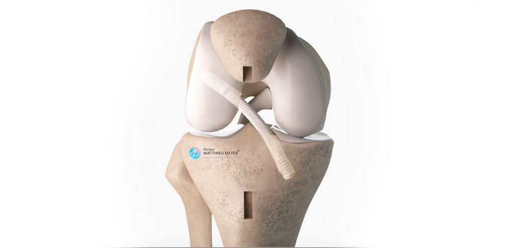
Revision Anterior Cruciate Ligament Reconstruction
In approximately 5% of cases, an anterior cruciate ligament ligamentoplasty will rupture and it is sometimes necessary to reconstruct the anterior cruciate ligament again. This iterative arthroscopic ligamentoplasty is more complex as it must take into account the tendon grafts available and the position of the bone tunnels created during the first ligamentoplasty. It is also recommended, if it was not done during the first ligamentoplasty, to combine the intra-articular reconstruction of the anterior cruciate ligament with an extra-articular plasty of the anterolateral ligament to reduce the risk of another ligament tear
Meniscal Surgery
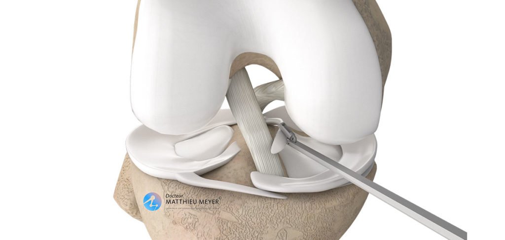
Arthroscopic Meniscal Surgery
Meniscus cracks are common. Most are degenerative due to the natural wear of the meniscus and others are caused by trauma. In some cases, surgery may be necessary. This is the case when a small piece comes away causing the knee to lock, in the event of meniscal damage in young subjects that can be repaired, or damage that is not relieved by medical treatments (injections). According to the age of the patient, as well as the type, shape, and location of the meniscal damage, the meniscus will be sutured or the damaged area removed. This operation is performed using arthroscopic surgery, a minimally invasive surgical technique.
Surgery to slow knee osteoarthritis
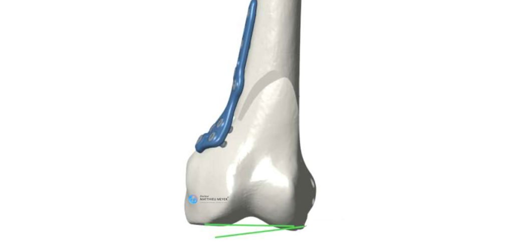
Medial Closing-Wedge Distal Femoral Osteotomy
A distal femoral (varus) osteotomy is the surgical realignment of the distal end of the femur which, in localized forms of gonarthrosis in young patients with genu valgum, reduces the mechanical load on the worn area of cartilage and transfers it to an area of healthy cartilage. The aim is to relieve the pain of gonarthrosis, slow the wear of the cartilage, and ultimately delay the need for a knee replacement for as long as possible.
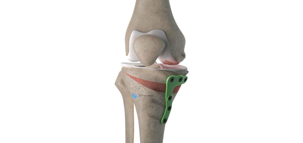
Medial Opening-Wedge High Tibial Osteotomy
A high tibial (valgus) osteotomy is the surgical realignment of the proximal end of the tibia which, in localized forms of gonarthrosis in young patients with genu varum, reduces the mechanical load on the worn area of cartilage and transfers it to an area of healthy cartilage. The aim is to relieve the pain caused by the gonarthrosis, slow the wear of the cartilage, and ultimately delay the need for a knee replacement for as long as possible.
Knee replacements
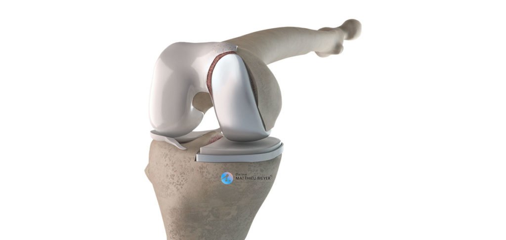
Partial Knee Replacement
A partial knee replacement is proposed when one of the two femorotibial compartments of the knee is worn and medical treatments no longer relieve the pain and the patient’s ability to get around has deteriorated. This operation aims to eliminate the pain and restore knee function so the patient can walk unrestrictedly and participate in gentle sports activities (walking, golf, cycling, swimming, keep-fit…). The minimally invasive technique used is gentle on the muscles and tendons and spares all the ligaments thus, in most cases, enabling faster recovery and a more natural feel than a total replacement.
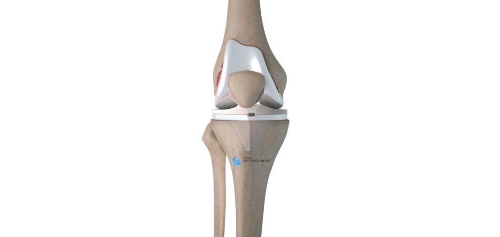
Total Knee Replacement
A total knee replacement is proposed in the case of diffuse wear of the knee cartilage when medical treatments no longer relieve the pain and the patient’s ability to get around has deteriorated. This operation aims to eliminate the pain and restore knee function so the patient can walk unrestrictedly and participate in gentle sports activities (walking, golf, cycling, swimming, keep-fit…).
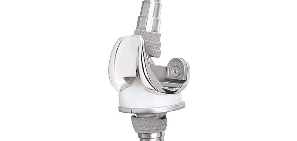
Revision Knee Replacement
In the case of wear, loosening or chronic infection of the knee implants, or wear of the remaining cartilage in the case of partial knee replacements, a revision knee replacement is necessary. This complex operation may require the use of a reconstruction prosthesis, creation of bone flaps or the use of bone grafts. The aim is to recover knee function and enable weight-bearing immediately on the limb operated on or later if bone grafts are required during the procedure.
Patellar instability surgery
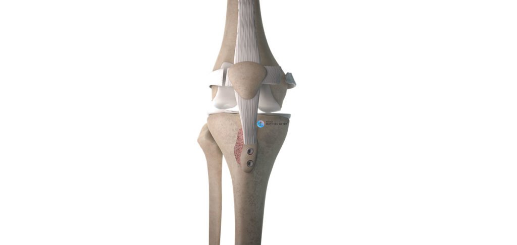
Anterior Tibial Tuberosity Transfer Patella Realignment
Patella realignment can be proposed in the case of recurrent dislocations of the patella or osteoarthritis of the lateral part of the femorotibial joint. Patella realignment involves the medial transfer of the anterior tibial tuberosity, a piece of bone located on the anterior side of the tibia in which the patellar tendon inserts. This is combined with cutting arthroscopically the patellar insertion of the vastus lateralis tendon, which helps decrease the lateral pulling forces and realign the patella with the femur.
Osteochondritis dissecans surgery
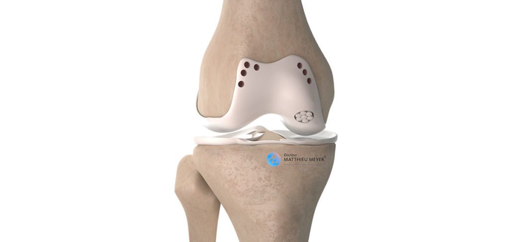
Drilling, Fixation And Osteochrondral Grafts
Osteochondritis dissecans can lead to the mobility and even partial or total detachment of a fragment of bone and cartilage from the femoral joint surface. According to the age of the patient, as well as the condition and mobility of the fragment, different surgical treatments are possible to relieve the pain and limit the risk of the subsequent onset of osteoarthritis. These treatments include deep cartilage drilling or reattachment of the mobile fragment, as well as more complex procedures such as bone and cartilage grafts or even grafts of cartilage cells grown in a laboratory.
Make an appointment
please do not hesitate to contact us or make an appointment online via DoctoLib
